Lily - Megaesophagus Secondary to PRAA
Meet Lily. Lily is an 8-week-old puppy with a history of chronic vomiting. The owners obtained Lily when she was 3 weeks old. As she aged, the owners tried to wean her to solid food. However, Lily would always regurgitate after eating. X-rays with and without barium were taken to verify a suspected megaesophagus and to determine if there was an area of narrowing (stricture).
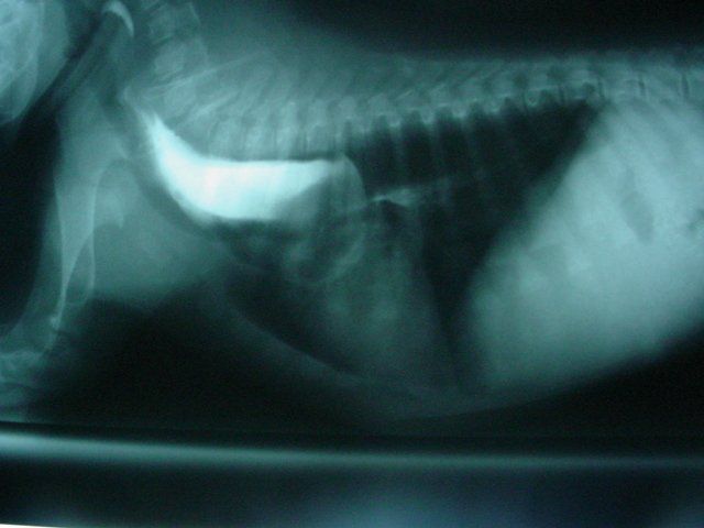
The x-ray below is one of the films taken with barium. Barium is a contrast media that can be used to outline hollow structures since it is bright white on x-rays. The x-ray on the left shows barium in a widened esophagus and a sudden area of narrowing and a lesser amount of barium after the stricture. The stricture occurs at the level of the 5th to 6th rib. In this area, it can be presumed that the stricture is caused by a persistent right aortic arch (PRAA) or vascular ring.
A PRAA is a blood vessel that should have deteriorated during normal development. In rare cases, it remains intact and can encircle the esophagus and prevent the passage of food into the stomach. As time goes on the esophagus ahead of the vessel will enlarge and overfill with food that is supposed to go to the stomach. Since the food cannot pass, the affected animal will regurgitate.
Early surgical intervention provides the best chance for long-term cure. Medical management will often be unsuccessful for megaesophagus due to PRAA. Inhalation of regurgitated food will often end in aspiration pneumonia and can be fatal. Lily's owners consulted about surgery.
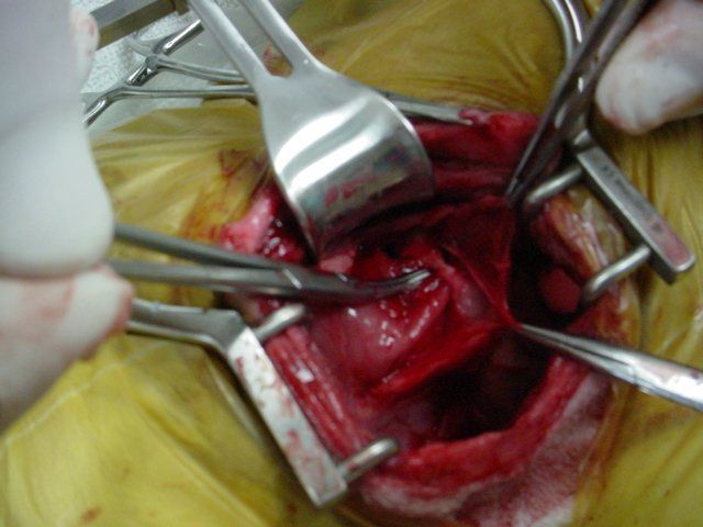
The picture below shows the isolated PRAA (under the curved tip clamp in the center). The enlarged portion of the esophagus is towards the left of the opening. The heart is towards the lower right of the picture. The opened mediastinum (tissues surrounding the heart and major vessels) is held within the two clamps on the right.
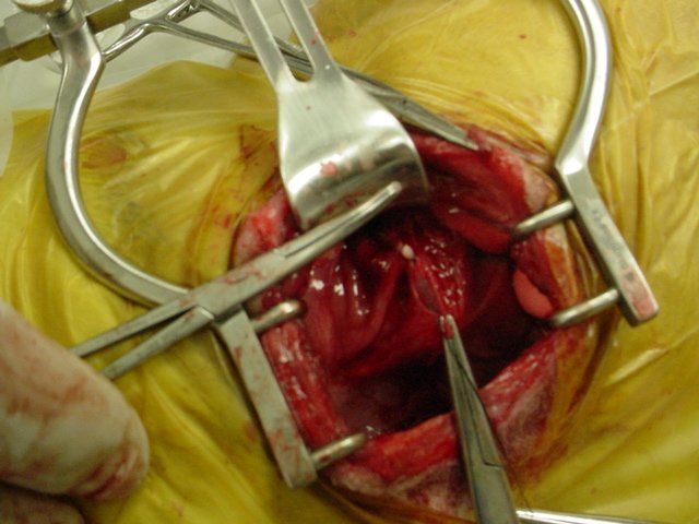
The vessel causing the entrapment (PRAA) was tied with two pieces of suture and cut. The picture below shows one end of the ligated vessel (small white circular tissue slightly above the lower clamp. The chest cavity was then closed in several layers. Lily will be fed small amounts of food several times per day for the next several weeks. With time, the enlarged portion of the esophagus should hopefully decrease in size and increase in contractility as the lower portion of the esophagus can now accept food. She will be monitored carefully for surgical complications and failure of the enlarged portion of the esophagus to regain normal function.
Learn More About
All Pets Veterinary Clinic

Share On: